
C-MORE POST
Post COVID-19 disease follow up imaging using hyperpolarised xenon MRI and CT
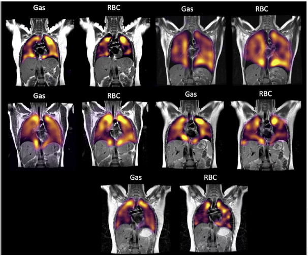
There is increasing clinical evidence that patients that have had severe COVID-19 pneumonia requiring hospitalisation remain severely out of breath on discharge and on follow up appointments. It is likely that COVID-19 infection may result in a more severe reduction of lung function, both because of its ability to induce respiratory distress and also potentially because of its tendency to produce blood clots in the lungs. This study aims to image the lungs of hospitalised and community-acquired COVID-19 patients using CT scans and Hyperpolarised Xenon MRI (HP 129Xe-MRI).
The unique ability of Xenon to dissolve through the respiratory membrane into the soft tissue of the lungs and capillaries may enable detection of much smaller changes in post COVID-19 pneumonia disease than has previously been possible when investigating changes in pulmonary function due to respiratory disease.
Combining the HP 129Xe-MRI images with the CT images will provide a unique insight into functional and structural changes in the lungs due to severe COVID-19 pneumonia. This research has gained national and global interest as it has been able to show clinical Long-COVID symptoms that traditional methods cannot detect.
XIPS
HP 129Xe-MRI in Idiopathic Pulmonary Fibrosis and Sarcoidosis
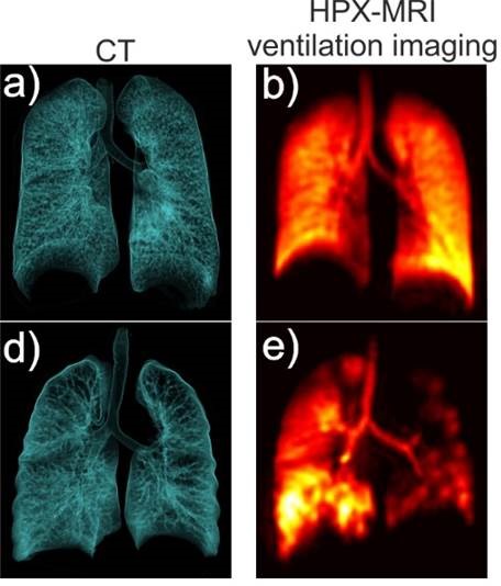
This new study aims to investigate the use of HP 129Xe-MRI as a method to detect changes in the integrity of lung tissue and blood vessels in patients with idiopathic pulmonary fibrosis (IPF). IPF is a disease characterised by progressive scarring of the lungs. Sarcoidosis is a multisystem inflammatory condition characterised by the formation of nodules at various sites in the body including the lung. The current tools of measuring these diseases and their response to medical treatment are relatively insensitive – CT scan interpretation is also subject to variability depending on who is viewing the scans. There is a need for a more sensitive and quantifiable method of measuring disease severity and change in order to improve the classification of IPF and sarcoidosis in patients and the monitoring of therapies. 129Xe is very soluble in lung blood and tissues which allow us to potentially measure very small changes in the lungs due to disease, in combination with anatomical lung imaging.
Xenon-Radiotherapy
Hyperpolarized Xenon gas MR Imaging in Radiotherapy
This study recruited participants who were receiving radiotherapy that affected the lungs. We wanted to obtain detailed images of lung structure as well as detailed airflow and blood flow throughout the lungs. Our aim was to see how sensitive the xenon images were over time – before, during and after treatment, in order to develop an objective and quantifiable method of lung assessment for these patients. The study has ended as of 2020 and a research paper will be published soon.

